A high magnification image of synapse obtained by electron microscopy
Por um escritor misterioso
Descrição

Large Volume Electron Microscopy and Neural Microcircuit Analysis
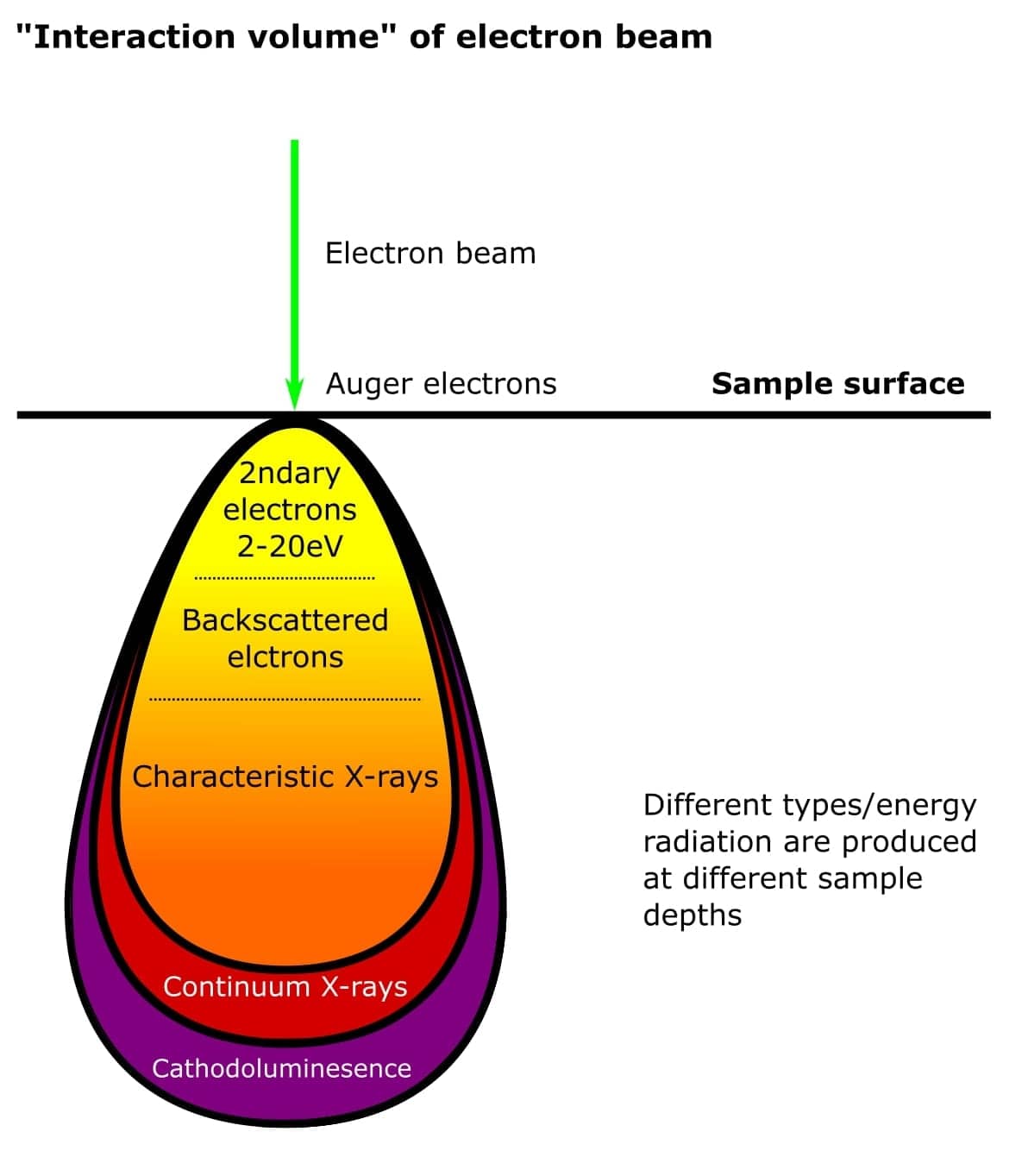
The 2 Main Electron Microscopy Techniques Explained

Auditory Hair Cell-Afferent Fiber Synapses Are Specialized to

Ultrastructural Imaging of Activity-Dependent Synaptic Membrane

High-resolution volumetric imaging constrains compartmental models

Modern field emission scanning electron microscopy provides new
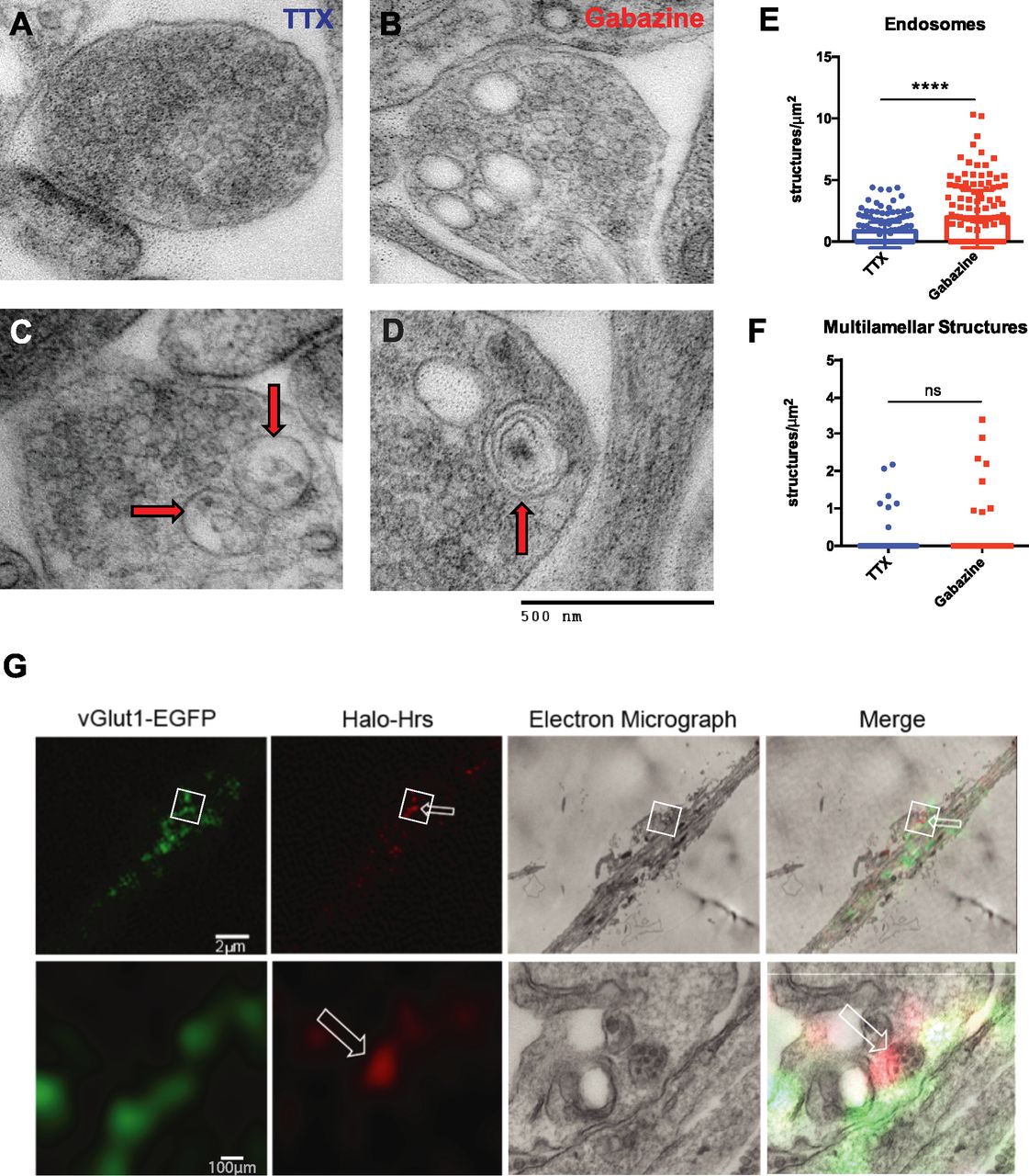
Axonal transport of Hrs is activity dependent and facilitates
An electron micrograph showing a typical synapse (arrow) within

What should we be measuring in brain preservation?
IJMS, Free Full-Text
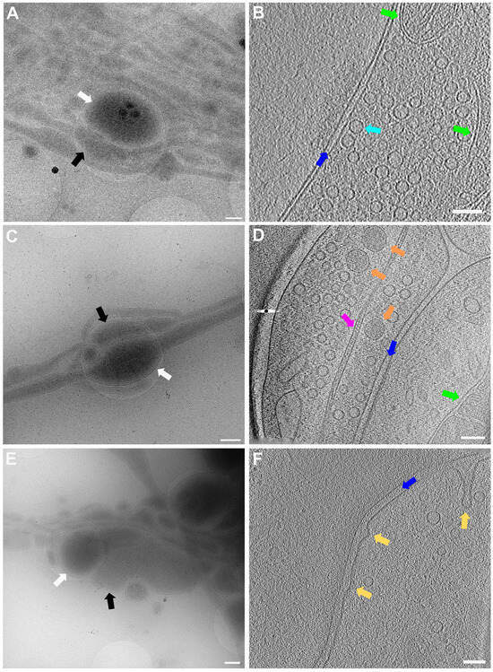
Light and electron microscopic imaging of synaptic vesicle
de
por adulto (o preço varia de acordo com o tamanho do grupo)