Figure 1 from Brain surface temperature under a craniotomy.
Por um escritor misterioso
Descrição
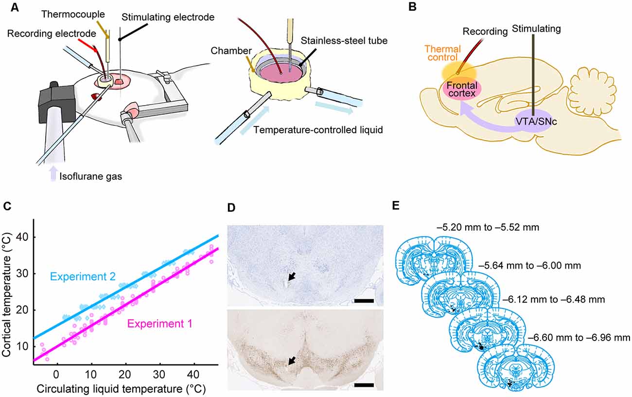
Fig. 1. Rapid cooling of the brain surface in an in vivo mouse preparation. A: schematic representation of a cranial window during recording of temperature and single-cell activity in the anesthetized mouse. The main potential routes of heat transfer are indicated. B: brain surface temperature measured with the thermocouple during replacement of the artificial cerebrospinal fluid (ACSF) with fresh ACSF warmed to 38°C. ACSF was replaced twice, indicated by the arrowheads. - "Brain surface temperature under a craniotomy."
tACS motor system effects can be caused by transcutaneous stimulation of peripheral nerves

Altered Cortical Trigeminal Fields Excitability by Spreading Depolarization Revealed with in Vivo Functional Ultrasound Imaging Combined with Electrophysiology

Reversible edema following electric drilling of macaque craniotomy

Cranial imaging window implantation technique for longitudinal multimodal imaging of the brain environment in live mice - ScienceDirect
A soft, transparent, freely accessible cranial window for chronic imaging and electrophysiology
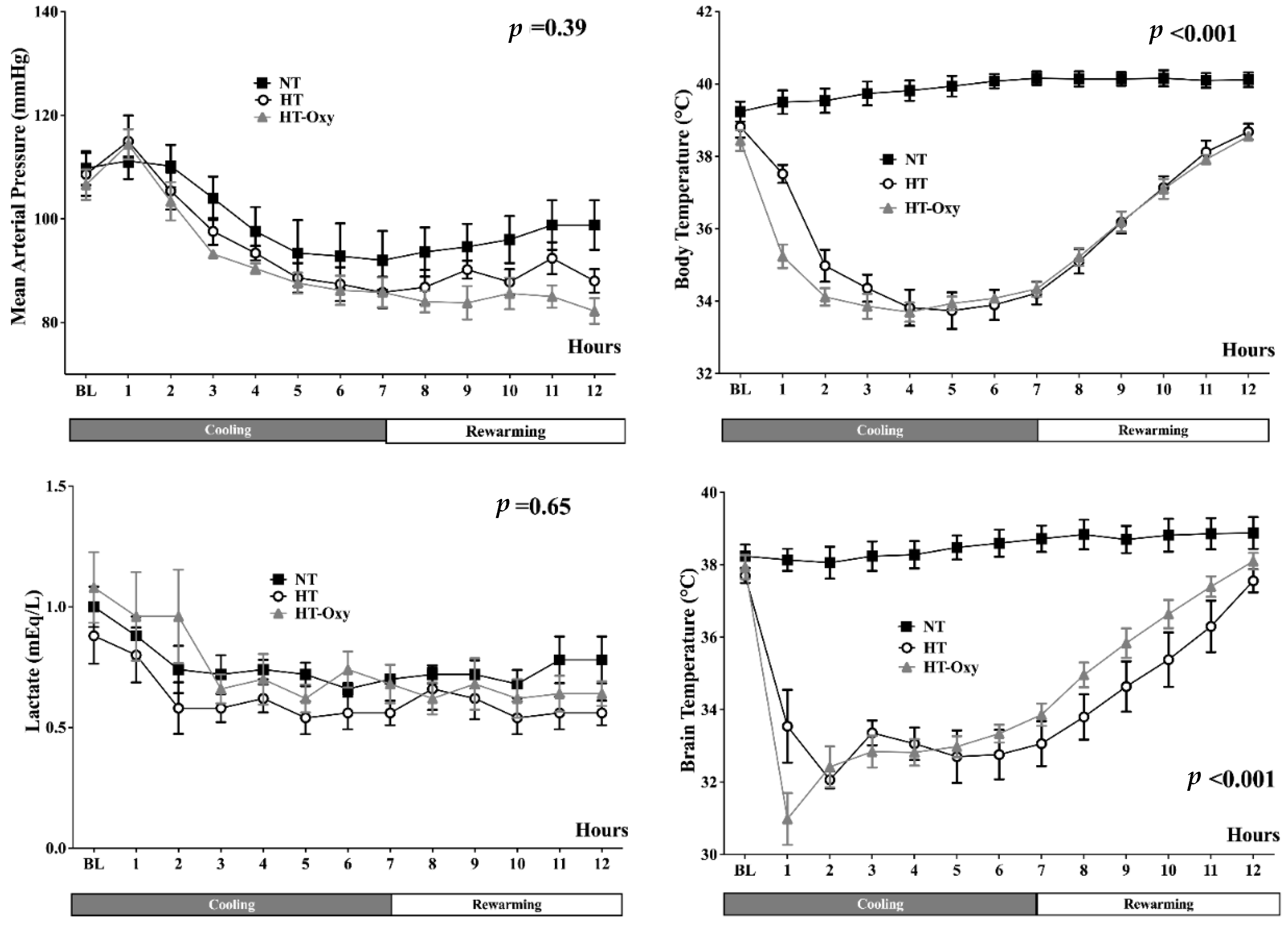
Frontiers Brain Temperature Alters Contributions of Excitatory and Inhibitory Inputs to Evoked Field Potentials in the Rat Frontal Cortex
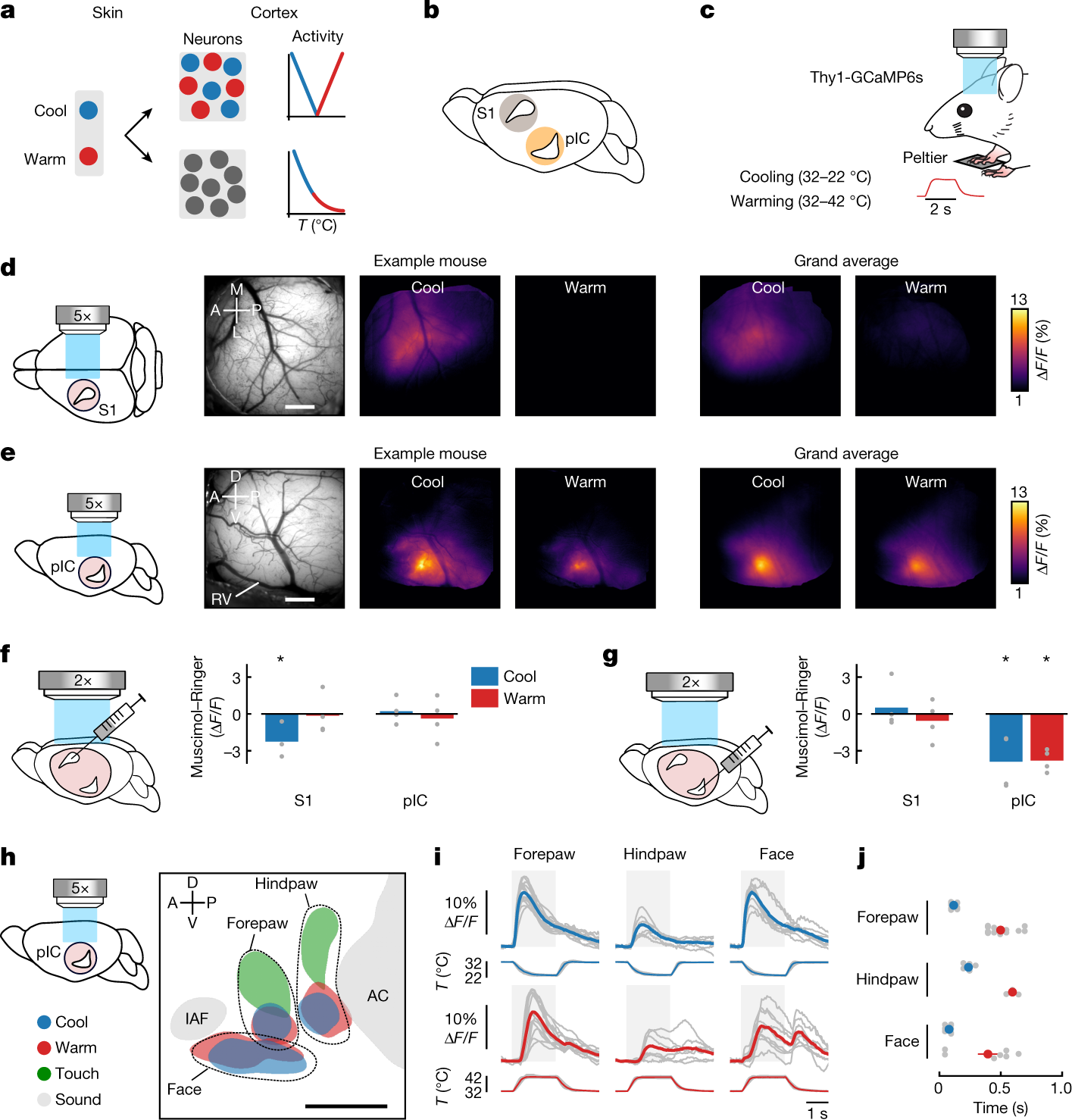
The cellular coding of temperature in the mammalian cortex
Brain Sciences, Free Full-Text
Brain Sciences, Free Full-Text
de
por adulto (o preço varia de acordo com o tamanho do grupo)